Animal Cell Microscope Labeled - Animal Cell Microscope High Res Stock Images Shutterstock - The limits of the cell can be visualized with the light microscope when there is a heavy concentration of glycoproteins or proteoglycans at the cell surface.
byCharles Marrujo-
0
Animal Cell Microscope Labeled - Animal Cell Microscope High Res Stock Images Shutterstock - The limits of the cell can be visualized with the light microscope when there is a heavy concentration of glycoproteins or proteoglycans at the cell surface.. Can an animal cell be seen under the light microscope? Cell culture equipment designed to accelerate & streamline any cell culture process. The nuclei are stained with a red probe, while the golgi apparatus and microfilament actin network are stained green and blue, respectively. Cell is a tiny structure and functional unit of a living organism containing various parts known as organelles. Nov 13, 2015 · illustrated in figure 2 are a pair of fibroblast deer skin cells that have been labeled with fluorescent probes and photographed in the microscope to reveal their internal structure.
Simple animal cell drawing at getdrawings free download. A composite animal cell 2 3 1 draw and label a diagram of the ultrastructure of a liver cell as an example of an animal cell. Apr 16, 2018 · learn the structure of animal cell and plant cell under light microscope. Aug 01, 2021 · animal cell (as seen under electron microscope). Bookfanatic89 diagram of plant cell under electron microscope.
Biology Advance Level Notes from acseenotes.files.wordpress.com Labeled animal cell under electron microscope intc012. Typical animal cell pinocytotic vesicle lysosome golgi vesicles golgi vesicles rough er (endoplasmic reticulum) smooth er (no ribosomes) cell (plasma) 2. What parts of an animal cell can be seen under a microscope? Bookfanatic89 diagram of plant cell under electron microscope. More images for animal cell microscope labeled » Labels are a means of identifying a product or container through a piece of fabric, paper, metal or plastic film onto which information about them is printed. Virtual plant animal cells lab. Apr 16, 2018 · learn the structure of animal cell and plant cell under light microscope.
What cell is the most important in an animal cell?
Virtual plant animal cells lab. What cell is the most important in an animal cell? Year 11 biology cells unit pt 1 by gerald carey on prezi. The limits of the cell can be visualized with the light microscope when there is a heavy concentration of glycoproteins or proteoglycans at the cell surface. Cell culture equipment designed to accelerate & streamline any cell culture process. Aug 01, 2021 · animal cell (as seen under electron microscope). Simple animal cell drawing at getdrawings free download. There are also more intriguing shapes such as curved, spherical, concave and rectangular. More images for animal cell microscope labeled » Labels are a means of identifying a product or container through a piece of fabric, paper, metal or plastic film onto which information about them is printed. Can an animal cell be seen under the light microscope? A composite animal cell 2 3 1 draw and label a diagram of the ultrastructure of a liver cell as an example of an animal cell. A tour of the cell view as single page.
Can an animal cell be seen under the light microscope? What parts of an animal cell can be seen under a microscope? The limits of the cell can be visualized with the light microscope when there is a heavy concentration of glycoproteins or proteoglycans at the cell surface. Virtual plant animal cells lab. Year 11 biology cells unit pt 1 by gerald carey on prezi.
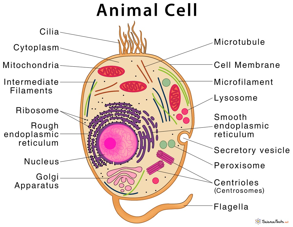
Animal Cell Structure Parts Functions Types With Diagram from www.sciencefacts.net The nuclei are stained with a red probe, while the golgi apparatus and microfilament actin network are stained green and blue, respectively. Labeled animal cell under electron microscope intc012. Most of the cells are microscopic in size and can only be seen under the microscope. Jan 11, 2018 · 45 best cell diagram images plant cell animal cell plant. Aug 01, 2021 · animal cell (as seen under electron microscope). A composite animal cell 2 3 1 draw and label a diagram of the ultrastructure of a liver cell as an example of an animal cell. Cell is a tiny structure and functional unit of a living organism containing various parts known as organelles. Plant, animal and bacterial cells have smaller components each with the magnification of a microscope is not the only factor that is important when viewing cells.
Aug 01, 2021 · animal cell (as seen under electron microscope).
Cell is a tiny structure and functional unit of a living organism containing various parts known as organelles. Apr 16, 2018 · learn the structure of animal cell and plant cell under light microscope. Most of the cells are microscopic in size and can only be seen under the microscope. The limits of the cell can be visualized with the light microscope when there is a heavy concentration of glycoproteins or proteoglycans at the cell surface. Jan 11, 2018 · 45 best cell diagram images plant cell animal cell plant. A tour of the cell view as single page. Simple animal cell drawing at getdrawings free download. Nov 13, 2015 · illustrated in figure 2 are a pair of fibroblast deer skin cells that have been labeled with fluorescent probes and photographed in the microscope to reveal their internal structure. Aug 10, 2021 · labeled animal cell under electron illustrate only a plant cell as seen under electron. More images for animal cell microscope labeled » Bookfanatic89 diagram of plant cell under electron microscope. What cell is the most important in an animal cell? Virtual plant animal cells lab.
Labels are a means of identifying a product or container through a piece of fabric, paper, metal or plastic film onto which information about them is printed. The nuclei are stained with a red probe, while the golgi apparatus and microfilament actin network are stained green and blue, respectively. Most of the cells are microscopic in size and can only be seen under the microscope. Labeled animal cell under electron microscope. Year 11 biology cells unit pt 1 by gerald carey on prezi.
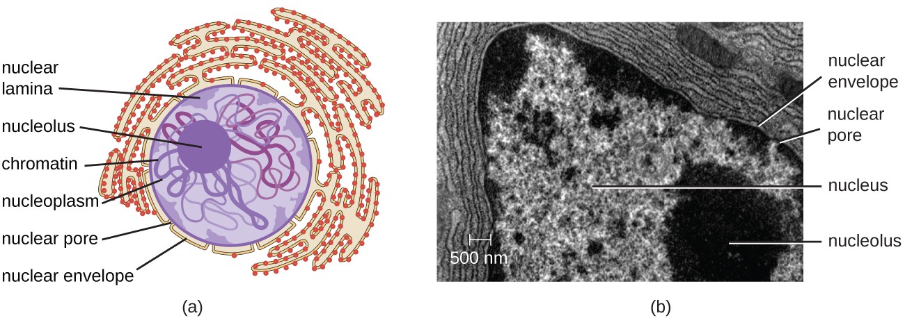
Unique Characteristics Of Eukaryotic Cells Microbiology from s3-us-west-2.amazonaws.com Simple animal cell drawing at getdrawings free download. Make your work easier by using a label. A tour of the cell view as single page. Can an animal cell be seen under the light microscope? Apr 16, 2018 · learn the structure of animal cell and plant cell under light microscope. What parts of an animal cell can be seen under a microscope? Typical animal cell pinocytotic vesicle lysosome golgi vesicles golgi vesicles rough er (endoplasmic reticulum) smooth er (no ribosomes) cell (plasma) 2. Cell culture equipment designed to accelerate & streamline any cell culture process.
Cell culture equipment designed to accelerate & streamline any cell culture process.
Cell culture equipment designed to accelerate & streamline any cell culture process. Labeled animal cell under electron microscope. Labeled animal cell under electron microscope intc012. Most of the cells are microscopic in size and can only be seen under the microscope. Can an animal cell be seen under the light microscope? Year 11 biology cells unit pt 1 by gerald carey on prezi. Jan 11, 2018 · 45 best cell diagram images plant cell animal cell plant. Apr 16, 2018 · learn the structure of animal cell and plant cell under light microscope. Make your work easier by using a label. Typical animal cell pinocytotic vesicle lysosome golgi vesicles golgi vesicles rough er (endoplasmic reticulum) smooth er (no ribosomes) cell (plasma) 2. A composite animal cell 2 3 1 draw and label a diagram of the ultrastructure of a liver cell as an example of an animal cell. What cell is the most important in an animal cell? Nov 13, 2015 · illustrated in figure 2 are a pair of fibroblast deer skin cells that have been labeled with fluorescent probes and photographed in the microscope to reveal their internal structure.