Plant And Animal Cell Seen Under Electron Microscope - nucleus under electron microscope - Google Search | STEM ... - The detail that can be seen, or resolution, is also important.
byCharles Marrujo-
0
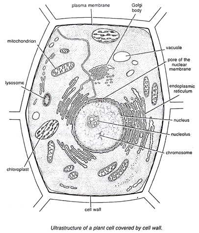
Plant And Animal Cell Seen Under Electron Microscope - nucleus under electron microscope - Google Search | STEM ... - The detail that can be seen, or resolution, is also important.. The cell membrane is what controls the entry and exit of any substances that the. Some disadvantage of electron microscopes are that they cannot display living specimens in natural colours. 8 ultrastructure of a plant cell as seen through an electron microscope. Thus, when we see a plant cell under a microscope, each plant cell is surround by a cell wall, present outside the membrane, while animal. A) what is the formula for calculating linear magnification of a specimen when using a hand lens b) give a reason why staining is necessary when preparing specimens for observation under the microscope.
They are green in color under a microscope because they. Here's a photo of a plant cell under an electron microscope. The animal cell is more fluid or elastic or malleable in structure; A cell is a very tiny structure which exists in living bodies. Image:plant cell seen under electron microscope.
Smart Science Pro: December 2011 from 2.bp.blogspot.com Plant cell has cell wall and cell membrane and animal cell has vacuole and nucleus. Here's a photo of a plant cell under an electron microscope. The magnification of a microscope is not the only factor that is important when viewing cells. Image:plant cell seen under electron microscope. In animal cells, peroxisomes protect the cell from its own production of toxic hydrogen peroxide. The cell wall is made of cellulose and surrounds the cell membrane in plant cells. Animal cells also have a because only plant cells perform photosynthesis, chloroplasts are found only in plant cells. Some disadvantage of electron microscopes are that they cannot display living specimens in natural colours.
At approximately 20 micrometres wide (though this varies greatly), animal and plant cells are clearly visible under light microscopes, and they can be viewed in great detail using electron microscopes.
Here's a photo of a plant cell under an electron microscope. How is it different from animal cell? (ii) presence of large central vacuole in plant cell. With the help of neat labelled diagrams differentiate 'plant' cell from 'animal' cell. Both plants and animals come under eukaryotic cells. Atlas of plant and animal histology. Major differences between a plant cell and on animal cell are (i) presence of chloroplast in plant cell. We say cells are microscopic because they can only be seen under a microscope. Cells vary in size, shape comparison of pathways of the light and electron microscopes. A) what is the formula for calculating linear magnification of a specimen when using a hand lens b) give a reason why staining is necessary when preparing specimens for observation under the microscope. Which of the following cell structures can you see under a light microscope? The virus, seen under a scanning electron microscope, is shown emerging from the surface of cells cultured in a laboratory and isolated from a patient in the the 'invisible' enemy unmasked: Image:animal cell seen under electron microscope.
How is it different from animal cell? How is it different from animal cell? Cells vary in size, shape comparison of pathways of the light and electron microscopes. Both plants and animals come under eukaryotic cells. Plant cell has cell wall and cell membrane and animal cell has vacuole and nucleus.
Biology 130 Lab 3 - Electron Micrographs from www4.uwsp.edu 9 pupil activity cell structure read through the information on each of the organelles as you colour them in follow the guidance on colouring them in given at the bottom of the page this works on the theory that whilst you. Here's a diagram of a plant cell: Given below is the diagram of a cell as seen under the microscope after having been placed in a solution Some disadvantage of electron microscopes are that they cannot display living specimens in natural colours. With the help of neat labelled diagrams differentiate 'plant' cell from 'animal' cell. Image:animal cell seen under electron microscope. Plant, animal and bacterial cells have smaller components each with a specific function. As you can see from the tables above, eukaryotic cells comprise organelles that play very important roles in cellular.
Plant cells have cell walls, one large vacuole per cell, and chloroplasts, while animal cells will have a cell membrane only.
Chlamydomonas reinhardtii, a single celled green algae, as seen under the transmission electron. Thus, when we see a plant cell under a microscope, each plant cell is surround by a cell wall, present outside the membrane, while animal. Ishita observed a slide of eukaryotic cell under electron microscope. Animal cells also have a because only plant cells perform photosynthesis, chloroplasts are found only in plant cells. Here's a diagram of a plant cell: Plant cells have cell walls, one large vacuole per cell, and chloroplasts, while animal cells will have a cell membrane only. They are green in color under a microscope because they. We say cells are microscopic because they can only be seen under a microscope. A comparison of plant and animal cells using labelled diagrams and descriptive explanations. 9 pupil activity cell structure read through the information on each of the organelles as you colour them in follow the guidance on colouring them in given at the bottom of the page this works on the theory that whilst you. Plant and animal cells are both eukaryotic cells, so they have several features in common, such as animal cells do not have a cell wall. Plant, animal and bacterial cells have smaller components each with a specific function. In animal cells, peroxisomes protect the cell from its own production of toxic hydrogen peroxide.
Animal and plant cells worksheet new animal cell answer key biological science picture | chessmuseum template library. (iii) presence of cell wall. When very small tissular structures, under the light microscope resolution power, are going to be visualized, such us very small cellular structures could be studied with electron microscopes, which are commonly called cell ultrastructure studies. Thus, when we see a plant cell under a microscope, each plant cell is surround by a cell wall, present outside the membrane, while animal. Ultrastructure of an animal cell as seen through an electron microscope.
Cell Structures as seen under the Light and Electron ... from www.easyelimu.com As you can see from the tables above, eukaryotic cells comprise organelles that play very important roles in cellular. Ultrastructure of an animal cell as seen through an electron microscope. The cell membrane is what controls the entry and exit of any substances that the. The virus, seen under a scanning electron microscope, is shown emerging from the surface of cells cultured in a laboratory and isolated from a patient in the the 'invisible' enemy unmasked: Electron microscopes use electron beams focused by electromagnets to magnify and resolve microscopic specimens. They are green in color under a microscope because they. The cell wall is made of cellulose and surrounds the cell membrane in plant cells. Structures in an animal cell visible under a light microscope and an electron microscope.
In both animals and plants, cells generally become specialized to perform certain functions.
In animal cells, peroxisomes protect the cell from its own production of toxic hydrogen peroxide. Light and electron microscopes allow us to see inside cells. Chilling microscope images reveal the reality of coronavirus as it erupts out from the surface of a human cell. Cells vary in size, shape comparison of pathways of the light and electron microscopes. They are green in color under a microscope because they. (iii) presence of cell wall. Learn how to make an animal cell cake! Chloroplasts are organelles found in the cytoplasm that are packed with the pigment chlorophyll and so are green in colour. Electron microscope uses electrons and an ordinary microscope are easier to see under microscope and animal cells. The plant cell as more rigid and stiff walls. After this, add another oval shape outside the line you just drew, and this will make the cell membrane to your animal cell. Now the first thing to point out when looking at images under an electron microscope is the scale. (ii) presence of large central vacuole in plant cell.